
Translate this page into:
Magnetic resonance imaging and intractable epilepsy: A systematic review
Address for correspondence: Dr. Fahad Alshehri, Department of Radiology, College of Medicine, Qassim University, Buraydah, Saudi Arabia. E-mail: f.alshehri@qu.edu.sa
This is an open-access article distributed under the terms of the Creative Commons Attribution-Noncommercial-Share Alike 3.0 Unported, which permits unrestricted use, distribution, and reproduction in any medium, provided the original work is properly cited.
This article was originally published by Qassim Uninversity and was migrated to Scientific Scholar after the change of Publisher.
Abstract
Objective:
Epilepsy is a chronic neurological disorder that occurs due to irregular neuronal activity in the central nervous system. The main job of a radiologist is to investigate the structural etiology in epilepsy patients. This study was undertaken to find out the importance of magnetic resonance imaging (MRI) in the screening of intractable epilepsy through a systematic search of literature.
Methods:
A systematic review was performed using the PRISMA guidelines. Peer-reviewed studies on MRI and intractable epilepsy were retrieved from MEDLINE, ScienceDirect and Google Scholar. Moreover, studies cited in the key articles were also screened to increase the sensitivity and specificity of the systematic search.
Results:
The database search till March 2022 found a total of 112610 articles. Out of them, only 10 highly selected articles were included in the study. The pooled data point out that the rapid development in MRI techniques and the functional MRI (fMRI) has now become more and more critical in the diagnosis and management of patients with epilepsy. In addition, the data also pointed out that MRI-based approaches are also very useful for post-operative epilepsy patients as it gives information about the quality of the surgery. The data collected showed that the MRI is the choice technique for the evaluation of patients with epilepsy.
Conclusions:
The applicability of MRI in epilepsy diagnosis is highly accessible in all over the globe. The pooled data concluded that the MRI-based surgical approaches are extremely useful for the surgeons to provide three-dimensional imaging with superimposed real-time pointer details that have proved successful for epilepsy patients.
Keywords
MRI
intractable epilepsy
systematic review
functional MRI

Introduction
Epilepsy is a chronic disorder of the central nervous system which resulted due to the abnormal activities of the neurons.[1] According to the recent reports, more than 70 million individuals have been affected with this neurological disorder which led to increase the global disease burden worldwide.[2] A number of epidemiological studies reported that the epilepsy is globally common but its prevalence is more in the developing countries as compared with its reported prevalence in developed countries and the situation to handle the epilepsy patients in low-income countries day-by-day becoming worst.[3] Several epidemiologist’s investigated that the prevalence rate of epilepsy has significantly increased in the rural areas as compared to the urban regions and this trend has almost the same in all over the globe.[4,5] Several investigators have investigated the reason(s) why the prevalence of epilepsy has higher in low-income countries or rural areas and they reported that the prevalence of epilepsy has direct correlation with the availability of the medical facilities such as qualified radiologists, surgeons, and also the availability of imaging techniques, which have been usually lacking in the low-income countries or in the rural areas.[6,7] The availability of the sophisticated technologies including the magnetic resonance imaging (MRI) has been found to be very useful for the management of patients with epilepsy in all over the world.[8,9] Imaging of epileptogenic lesions through MRI not only provides the planning for the surgeons to perform epilepsy surgeries but also helpful for the post-operative follow-up.[10] According to the International League Against Epilepsy (ILAE) suggests that all individuals suffering from epilepsy ideally tested with high-quality imaging techniques including MRI.[11] Moreover, the National Institute of Health and Clinical Excellence (NICE) guidelines recommended that the structural abnormalities in the brain of children and adults suffering with epilepsy should be screened by MRI.[11,12] In modern world, functional MRI (fMRI) has now becomes the method of choice for the screening of epileptogenic lesions in the brain of individuals suffering from epilepsy as it is more powerful, highly sensitive, and more accurate as compared with the conventional MRI technique.[12,13] But still, the facility of fMRI in the rural regions or in low-income countries is not fully available due to the financials reasons, therefore, the radiologists working in these regions is still relies on the conventional MRI.[14,15] Under such circumstances, the radiologists play a key role to determine and to locate the structural abnormality of epileptogenic lesions to provide an accurate information to the surgeons so that the surgeon can easily identify the epileptogenic lesions and perform the surgeries.[16,17] In view of these, this systematic review was designed to update the role of the MRI in the screening of epilepsy by pooling of the available literature. The outcome of the pooling of selected studies clearly indicating that the MRI-based surgical systems provide a three-dimensional pictorial presentation of anatomic epileptogenic lesions with superimposed real-time pointer information to the surgeons that proved to be useful in epilepsy surgery. In general, the MRI-based therapies are extremely valuable for the management of patients with epilepsy.
Methods
PRISMA guidelines and the databases for systematic search of literature
The PRISMA guidelines part for a systematic search of literature was used in this systematic review.[18] A systematic computerized search using the databases such as PubMed, ScienceDirect, and Google Scholar was used. We identified studies that reported data on the role of magnetic resonance imaging in intractable epilepsy. The articles we use include all types of studies ranging from case reports, observational studies, randomized controlled trials, systemic reviews, and meta-analysis. Any patient, including male, female, transgender, any ethnic, and from any location, can be included in this study. A study of the abstracts of the chosen articles was performed, and only those with data deemed suitable for review were shortlisted. However, the references found to be relevant were also assessed and reviewed to be included in this study. The following keywords were used for our regular search: Magnetic resonance imaging and intractable epilepsy. Only English language studies were included to avoid misinterpretation of translation. All the data used in this literature review are collected ethically and legally. The search method is summarized as a PRISMA flowchart in Figure 1.
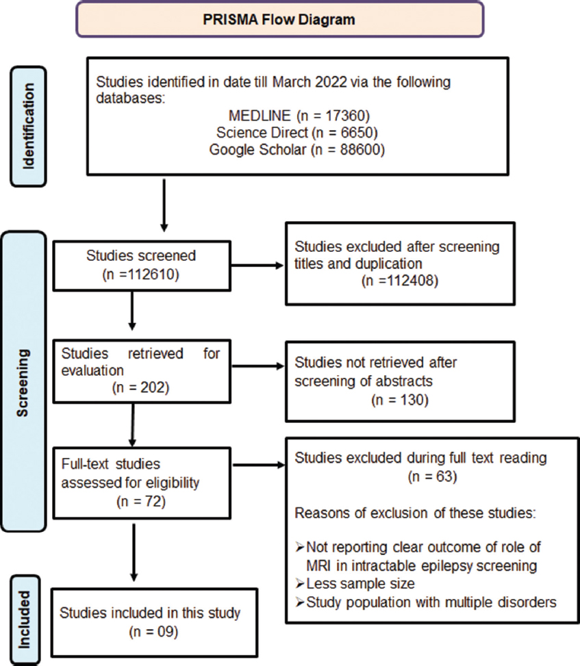
- PRISMA flowchart for systematic literature search
Inclusion and exclusion criteria of the studies
We included all retrospective and prospective population-based studies measuring MRI in epilepsy. We considered studies for inclusion if they included a definition of MRI and epilepsy. We excluded studies if they explored only epilepsy. We excluded editorials, single cases, and case series, studies published only as abstracts, letters, or commentaries, studies of individual groups.
Ethical issues
This is a systematic literature review, and the information of patients directly obtained from the published literature. Therefore, ethical approval was not required.
Results
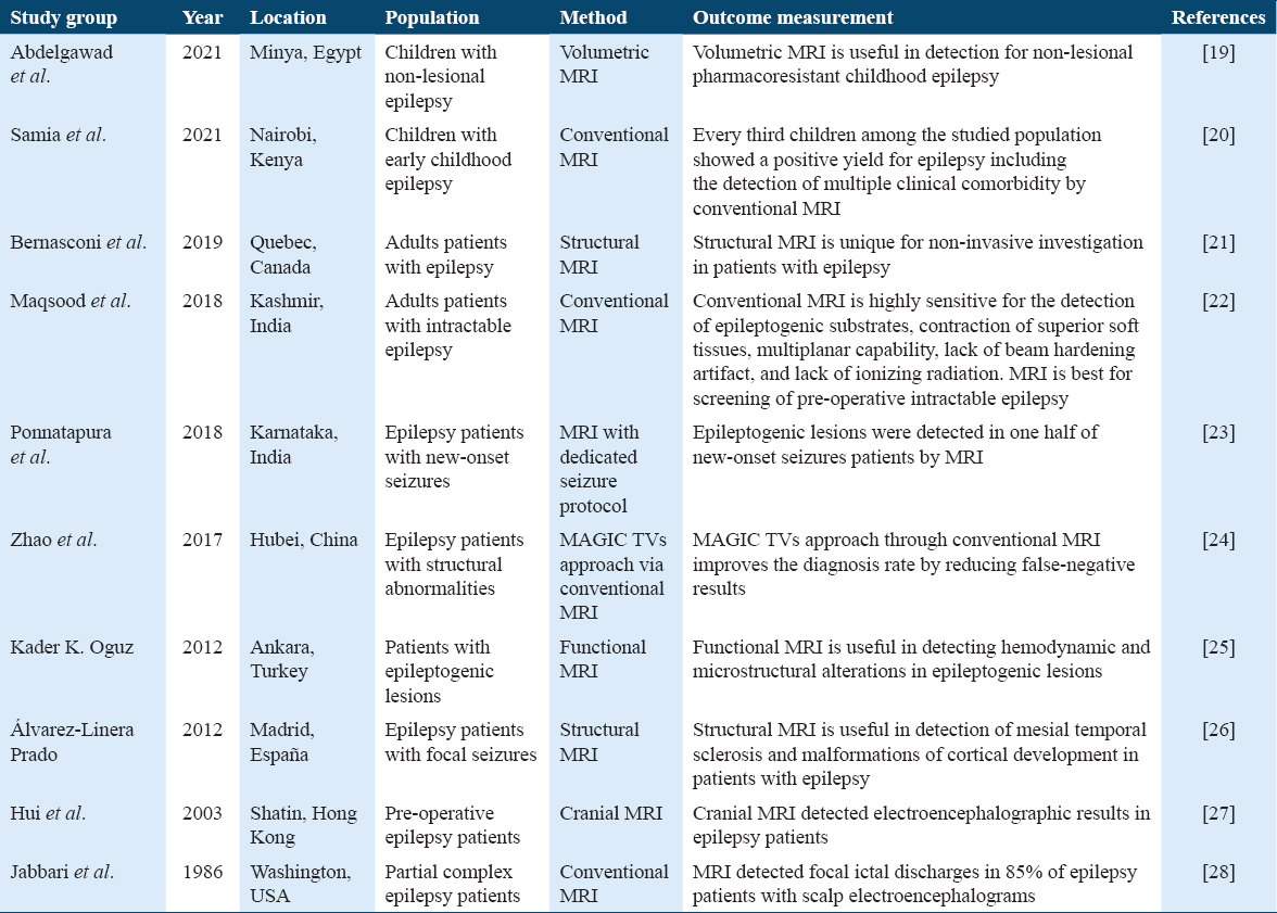
In this systematic review, our database search till March 2022 using the keywords MRI and epilepsy found 17360 articles on MEDLINE, 6650 articles on ScienceDirect, and 88,600 articles on Google Scholar found 112,610 articles. Out of them, only 202 articles were retrieved and rest were excluded after screening of titles and duplication. Out of 202 selected articles, 130 articles were excluded based on their relevancy of the abstract. Afterward, 72 articles were shortlisted and their full texts were accessed. After analyzing the full texts, 63 articles were excluded as these articles were not reported a clear outcome of the role of MRI in intractable epilepsy, few were excluded as their outcomes were based on significantly less sample size. The articles reported that the same previously published data were also excluded [Figure 1]. A total of 10 highly selective studies were included in this article.[19-28] A study conducted on children with non-lesional epilepsy from Minya, Egypt, in 2021 by Abdelgawad et al. who showed that volumetric MRI is extremely useful in the analysis of non-lesional pharmacoresistant childhood epilepsy.[19] In 2021, another published study on the Kenyan children with early childhood epilepsy reported that the conventional MRI is useful for the detection a multiple clinical comorbidity in childhood epilepsy.[20] In 2019, Bernasconi et al. from Quebec, Canada, showed that the structural MRI is very useful for the detection of noninvasive lesions in patients with epilepsy.[21] Another interestingly study from Jammu Kashmir, India, pointed out that the conventional MRI is highly sensitive for the detection of epileptogenic materials, dysfunctionality in soft tissues and they reported that MRI is the best technique for the screening of pre-operative intractable epilepsy.[22] Another study from Karnataka, India, showed that MRI with dedicated seizure protocol is best for the detection of epileptogenic lesions in one half of the new-onset seizures in epilepsy patients.[23] An interestingly study from Hubei, China, by Zhao et al. in 2017 showed that Magic TVs approach through conventional MRI significantly improved the diagnosis of epilepsy by reducing the outputs of negative findings.[24] Kader K Oquz from Ankara, Turkey, showed that functional MRI is an important technique for the detection of hemodynamic and microstructural alterations in epileptogenic lesions.[25] In 2012, a study from Madrid, España, conducted on the epilepsy patients with focal seizures reported that the structural MRI technique is useful in the detection of mesial temporal sclerosis and malformations of cortical development in epilepsy patients.[26] Hui et al. from Hong Kong pointed out that the cranial MRI technique is useful to show electroencephalographic results in patients with pre-operative epilepsy.[27] A study from Washington DC, USA, by Jabbari et al. investigated that the conventional MRI has potential to detect focal ictal discharges in up to 85% of epilepsy patients with scalp electroencephalograms.[28] The detailed characteristics with their outcomes are summarized in Table 1. In short, the applicability of MRI in epilepsy diagnosis is highly accessible to most of the population in developed economies. With the rapid development of MRI, exceptionally functional MRI (fMRI) is becoming more and more critical in the diagnosis and management of patients with epilepsy. The outcomes of this systematic review clearly point out that the MRI is choice technique for the evaluation of patients with epilepsy.
Discussion
This systematic review is provided an updated knowledge on the role of the MRI in the screening of structural abnormalities of the neural lesions in patients with epilepsy. The MRI examination plays a key role for the screening of epileptogenic lesions in patients with epilepsy, this may be conducted either through MRI technique alone or in combination with some other imaging techniques such as fMRI, MR spectroscopy, PET, and ictal SPECT.[29] The performance of medical examination of epileptogenic lesions with these additional approaches provides an extremely valuable information about the epileptogenic lesions and its exact location in the brain, these additional approaches with MRI not only help the surgeons to perform epileptogenic lesional surgery but also help the radiologists to make their outstanding reports.[30,31] In spite of the availability of these extraordinary functional imaging approaches but still the applicability of these approaches are limited.[30] There have been several reasons but the most valid reason is the involvement cost in these techniques while applying for the analysis of anatomic epileptogenic lesions.[30,31] Several investigators have reviewed all these additional techniques with MRI and all have concluded that these additional techniques are highly sensitive, more powerful, and accurate when performed with MRI.[31] Applicability of these functional imaging techniques is not the same at all time but these have been usually implemented in their specific requirements for the screening of epilepsy such as electroencephalogram (EEG) which is extremely useful for the detection of epileptogenic lesions in the brain when it can be recorded during fMRI.[32] Whereas, PET is highly useful for the detection of cortical dysplasia, which usually has not been detected by the conventional MRI technique in patients with epilepsy.[33] Furthermore, epilepsy patients have also been screened by MRI-based invasive EEG monitoring and also through placing of intracranial depth recording electrodes.[34,35] These approaches have provided a top level screening for the detection of highly complicated epileptogenic lesions.[34,35] In addition, MRI-based screening guidelines for the surgeons have also been developed and implemented which provided three-dimensional knowledge with superimposed real-time details of epileptogenic lesions to the surgeons which have already been proven to be successful for the performing epilepsy surgeries.[24-26] Not only have these, the post-operative MRI has also provided an information for the quality of surgery, whether it can be done in a correct manner and it also provides information whether the second round surgery should be needed.[33] Further, almost all types of brain-associated major structural abnormalities including cortical resection, corpus callosotomy, and hemispherectomy have been screened and analyzed by MRI.[33-35] Therefore, MRI application either alone or in combination with other functional approaches is highly applicable for the diagnosis of epilepsy patients. In this systematic review, we have provided the role of various types of MRIs for the screening of patients of different stages of intractable epilepsy. Using the keywords MRI and epilepsy, our systematic searched of literature initially found a total of 112,610 articles on MEDLINE, ScienceDirect, and Google Scholar. After reviewing studies title, abstracts, and full text at different stages, only 10 highly relevant studies were included in this study. In 2021, a study conducted by Abdelgawad et al.[19] on children with non-lesional epilepsy in Minya, Egypt, reported that volumetric MRI is extremely useful in the analysis of non-lesional pharmacoresistant childhood epilepsy. In another study, Samia et al. from Nairobi, Kenya, reported that the conventional MRI has potential to detect multiple clinical comorbidity in childhood epilepsy.[20] Furthermore, Bernasconi et al. from Quebec, Canada, in 2019 reported that the structural MRI is very useful for the detection of non-invasive lesions in patients with epilepsy.[21] Importantly, a study by Maqsood et al. from Jammu Kashmir, India, pointed out that the conventional MRI is highly sensitive for the detection of epileptogenic materials, dysfunctionality in soft tissues and they reported that MRI is the best technique for the screening of pre-operative intractable epilepsy.[22] Moreover, Ponnatapura et al. from Karnataka, India, in 2018 reported that MRI with dedicated seizure protocol is best for the detection of epileptogenic lesions in one half of the new-onset seizures in epilepsy patients.[23] Furthermore, Zhao et al. from Hubei, China, reported that Magic TVs approach through conventional MRI improves the diagnosis of epilepsy by reducing negative outcomes.[24] In another study, functional MRI found to be an important technique for the detection of hemodynamic and microstructural alterations in epileptogenic lesions.[25] In addition, another study showed that the structural MRI technique has potential for the analysis of mesial temporal sclerosis and malformations of cortical development in epilepsy patients.[26] In another study, cranial MRI technique was found to be useful for presenting findings in electroencephalographs in patients with pre-operative epilepsy.[27] Moreover, conventional MRI was also found to be an important for the detection of focal ictal discharges in patients with scalp electroencephalograms epilepsy.[28] All these data clearly pointed out that all types of MRI whether perform alone or in combinations with other techniques are extremely useful for the screening of intractable epilepsy.
Conclusions
The applicability of magnetic resonance imaging for the diagnosis of epilepsy is highly accessible in all over the globe. With the advancement in the MRI technique, this can be also applying with additional approaches. The functional MRI has now becoming more and more critical in the diagnosis and management of highly complicated epilepsy patients. The outcomes of this systematic review clearly point out that the MRI is a choice technique for the evaluation of epileptogenic lesions. The pooling of selected studies clearly indicating that the MRI-based surgical approaches provide three-dimensional pictorial details with superimposed real-time surgeons pointer information for the epilepsy patients, which already been proved successful in epilepsy surgery. In addition, the data also pointed out that MRI-based approach is also very useful for post-operative epilepsy patients as it gives information about the quality of the surgery. In short, the MRI-based therapies are extremely valuable for the management of patients with epilepsy.
Authors’ Declaration Statements
Availability of data and material
The data used in this study are available and will be provided by the corresponding author on a reasonable request.
Competing interests
None.
ORCID link of the submitting author: https://orcid.org/0000-0001-5755-7350
References
- Epidemiological profile of epilepsy in low income populations. Seizure. 2018;56:67-72.
- [Google Scholar]
- The global burden of epilepsy report:Implications for low- and middle-income countries. Epilepsy Behav. 2020;105:106949.
- [Google Scholar]
- Epidemiology, aetiology, and clinical management of epilepsy in Asia:A systematic review. Lancet Neurol. 2007;6:533-43.
- [Google Scholar]
- Epilepsy in Asia:Disease burden, management barriers, and challenges. Epilepsia. 2019;60:7-21.
- [Google Scholar]
- Packages of care for epilepsy in low- and middle-income countries. PLoS Med. 2009;6:e1000162.
- [Google Scholar]
- Identification of the epileptic focus:Magnetic resonance imaging. Epilepsy Res Suppl. 1992;5:95-100.
- [Google Scholar]
- Operational classification of seizure types by the international league against Epilepsy:Position paper of the ILAE commission for classification and terminology. Epilepsia. 2017;58:522-30.
- [Google Scholar]
- Standard magnetic resonance imaging is inadequate for patients with refractory focal epilepsy. J Neurol Neurosurg Psychiatry. 2002;73:643-7.
- [Google Scholar]
- 3 TESLA MR imaging in adults with focal onset epilepsy. Clin Neurol Neurosurg. 2013;115:2111-6.
- [Google Scholar]
- Contribution of MRI to the exploration of partial refractory epilepsy. Rev Neurol (Paris). 2004;160:5S91-7.
- [Google Scholar]
- High-resolution MRI enhances identification of lesions amenable to surgical therapy in children with intractable epilepsy. Epilepsia. 2004;45:954-9.
- [Google Scholar]
- ILAE classification of the epilepsies:Position paper of the ILAE commission for classification and terminology. Epilepsia. 2017;58:512-21.
- [Google Scholar]
- The PRISMA statement for reporting systematic reviews and meta-analyses of studies that evaluate healthcare interventions:Explanation and elaboration. BMJ. 2009;339:b2700.
- [Google Scholar]
- Magnetic resonance imaging (MRI) volumetry in children with nonlesional epilepsy, does it help? Egypt J Radiol Nucl Med. 2021;52:35.
- [Google Scholar]
- Magnetic resonance imaging findings in childhood epilepsy at a tertiary hospital in Kenya. Front Neurol. 2021;12:623960.
- [Google Scholar]
- Recommendations for the use of structural magnetic resonance imaging in the care of patients with epilepsy:A consensus report from the international league against epilepsy neuroimaging task force. Epilepsia. 2019;60:1054-68.
- [Google Scholar]
- Utility of magnetic resonance imaging brain epilepsy protocol in new-onset seizures:How is it different in developing countries? J Clin Imaging Sci. 2018;8:43.
- [Google Scholar]
- Role of conventional magnetic resonance imaging in the screening of epilepsy with structural abnormalities:A pictorial essay. Am J Nucl Med Mol Imaging. 2017;7:126-37.
- [Google Scholar]
- Role of magnetic resonance imaging for preoperative evaluation of patients with refractory epilepsy. Hong Kong Med J. 2003;9:20-4.
- [Google Scholar]
- Magnetic resonance imaging in partial complex epilepsy. Arch Neurol. 1986;43:869-72.
- [Google Scholar]
- Long-term epilepsy surgery outcomes in patients with MRI-negative temporal lobe epilepsy. Epilepsia. 2010;51:2260-9.
- [Google Scholar]
- Imaging intractable epilepsy:How many tests are enough? Am J Neuroradiol. 1999;20:534-5.
- [Google Scholar]
- The relative contributions of MRI, SPECT, and PET imaging in epilepsy. Epilepsia. 1994;35:S72-89.
- [Google Scholar]
- Acquiring simultaneous EEG and functional MRI. Clin Neurophysiol. 2000;111:1974-80.
- [Google Scholar]
- Comparison of MR imaging with PET and ictal SPECT in 118 patients with intractable epilepsy. AJNR Am J Neuroradiol. 1999;20:593-9.
- [Google Scholar]
- Magnetic resonance imaging:Principles and techniques:Lessons for clinicians. J Clin Exp Hepatol. 2015;5:246-55.
- [Google Scholar]
- Accessible magnetic resonance imaging:A review. J Magn Reson Imaging. 2019;49:e65-77.
- [Google Scholar]


